AUTHOR OF THIS BLOG

DR ANTHONY MELVIN CRASTO, WORLDDRUGTRACKER

Trimyristin

 PROCESS, spectroscopy  Comments Off on Trimyristin
Jul 122015
 

Trimyristin

Trimyristin

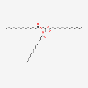
Trimyristin is an ester with the chemical formula C45H86O6. It is a saturated fat which is the triglyceride of myristic acid. Trimyristin is a white to yellowish-gray solid that is insoluble in water, but soluble in ethanol, benzene, chloroform, dichloromethane, and ether.


Name Trimyristin
Synonyms Glycerol trimyristate
Name in Chemical Abstracts Tetradecanoic acid, 1,2,3-propanetriyl ester
CAS No 555-45-3
EINECS No 209-099-7
Molecular formula C45H86O6
Molecular mass 723.18
SMILES code CCCCCCCCCCCCCC(=O)OCC(OC(=O)CCCCCCCCCCCCC)COC(=O)CCCCCCCCCCCCC
Nutmeg
reacts to
Trimyristin

TRIMYRISTIN.png

Occurrence

Trimyristin is found naturally in many vegetable fats and oils.

Isolation from nutmeg

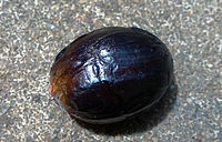
Seed of nutmeg contains trimyristin

The isolation of trimyristin from powdered nutmeg is a common introductory-level college organic chemistry experiment. It is an uncommonly simple natural product extraction because nutmeg oil generally consists of over eighty percent trimyristin. Trimyristin makes up between 20-25% of the overall mass of dried, ground nutmeg. Separation is generally carried out by steam distillation and purification uses extraction from ether followed by distillation or rotary evaporation to remove the volatile solvent. The extraction of trimyristin can also be done with diethyl ether at room temperature, due to its high solubility in the ether. The experiment is frequently included in curricula, both for its relative ease and to provide instruction in these techniques.

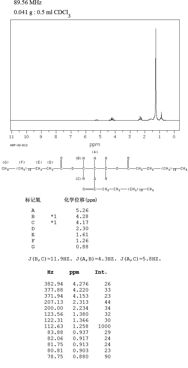
1H-NMR

1H NMR

1H-NMR: Trimyristin
300 MHz, CDCl3
delta [ppm] mult. atoms assignment
0.90 m 9 H 14-H (CH3)
1.2-1.4 m 60 H 4-13-H (CH2)
1.5-1.7 m 6 H 3-H
2.33 m 6 H 2-H
4.16 dd 2 H glycerol-C1-Ha
4.31 dd 2 H glycerol-C1-Hb
5.28 m 1 H glycerol-C2-H
7.26 CHCl3
2.11 acetone (impurity)

Isolation of trimyristin from nutmeg

Reaction type: isolation of natural products
Substance classes: carboxylic acid ester, triglyceride, natural product
Techniques: extracting with Soxhlet extractor, evaporating with rotary evaporator, recrystallizing, filtering, heating under reflux, heating with oil bath, stirring with magnetic stir bar
Degree of difficulty: Easy
Batch scale: 25 g Nutmeg

Reaction……….http://kriemhild.uft.uni-bremen.de/nop/en/instructions/pdf/1021_en.pdf

The reaction apparatus consists of a 250 mL round-bottom flask with a magnetic stir bar and a 100 mL soxhlet extraction unit with a reflux condenser. 25 g of finely ground nutmeg are placed into the extraction sleeve and covered with a little glass wool. 150 mL tert-butyl methyl ether are placed into the flask and whilst stirring, the solvent is heated to reflux until the solvent leaving the extraction sleeve is colourless (approximately 5 hours).

Work up

The solvent is evaporated with a final pressure of 20 hPa. The flask containing the residue is cooled in an ice bath or the refrigerator until the contents has crystallized to a thick slurry.

Crude product yield: 12 g;

The crude product is recrystallized from the minimum amount of ethanol. Prior to filtering the crystals, the flask is placed into the refrigerator for at least 30 minutes. The crystalline slurry is filtered and the product is dried in an evacuated desiccator over silica gel. Should the crystals not be colourless after the first recrystallization, a second recrystallization is carried out.

Yield: 6.5 g; melting point 54-55 °C;

Duration of the experiment

Without recrystallization 6 hours

Where can I stop the experiment?

Before and after the evaporation of the solvent

Recycling

The evaporated tert-butyl methyl ether and the evaporated ethanol from the mother liquor are collected and redistilled.

Suggestions for waste disposal

Waste Disposal
residue from mother liquor domestic waste
residue from extraction domestic waste

Operating scheme

Operating scheme

Substances required

Batch scale: 25 g Nutmeg
Educts Amount Risk Safety
Nutmeg
25 g R S
Solvents Amount Risk Safety
Ethanol
F F
~ 150 mL R 11 S 2-7-16
tert-Butyl methyl ether
F F Xi Xi
150 mL R 11-38 S 2-9-16-24
Others Amount Risk Safety
Iodine
Xn Xn N N
0.1 g R 20/21-50 S 2-23.2-23.4-25-61
Solvents for analysis Amount Risk Safety
Cyclohexane
F F Xn Xn N N
? R 11-38-50/53-65-67 S 2-9-16-33-60-61-62
Acetic acid ethyl ester
F F Xi Xi
? R 11-36-66-67 S 2-16-26-33

Substances produced

Batch scale: 25 g Nutmeg
Products Amount Risk Safety
Trimyristin
6.5 g R S

Equipment

Batch scale: 25 g Nutmeg
round bottom flask 250 mL round bottom flask 250 mL Soxhlet extractor 100 mL Soxhlet extractor 100 mL
glass wool glass wool extraction cone extraction cone
heatable magnetic stirrer with magnetic stir bar heatable magnetic stirrer with magnetic stir bar oil bath oil bath
reflux condenser reflux condenser rotary evaporator rotary evaporator
ice bath ice bath exsiccator with drying agent exsiccator with drying agent
suction filter suction filter suction flask suction flask

Simple evaluation indices

Batch scale: 25 g Nutmeg
Atom economy not defined
Yield not defined
Target product mass 6.5 g
Sum of input masses 250 g
Mass efficiency 26 mg/g
Mass index 39 g input / g product
E factor 38 g waste / g product
Energy input 1500 kJ
Energy efficiency 4.3 mg/kJ

Chromatogram

crude product chromatogram

TLC: crude product
TLC layer Polygram SilG/UV precoated TLC layer; 0.2 mm; silica gel; Macherey & Nagel
mobile phase cyclohexane / EtOAc = 95 : 5
staining reagent Vaughn’s reagent or iodine vapor
Rf (product) 0.51

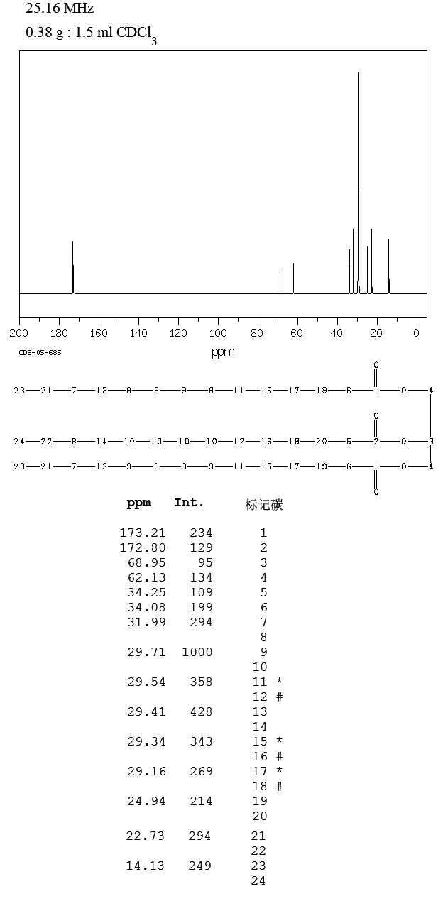
13C-NMR

13C NMR

13C-NMR: Trimyristin
300 MHz, CDCl3
delta [ppm] assignment
14.08 C14
22.66 C13
24.85-24.89 C3, C17
29.06-31.90 C4-C12
34.04-34.2 C2
62.08 glycerol-C1
68.85 glycerol-C2
172.85 C15
173.26 C1
76.5-77.5 CDCl3

IR

IR

IR: Trimyristin
[KBr, T%, cm-1]
[cm-1] assignment
2950-2850 aliph. C-H valence
1730 C=O valence, ester


Trimyristin[1]
Skeletal formula of trimyristin
Ball-and-stick model of trimyristin
Space-filling model of trimyristin
Names
IUPAC name

1,3-Di(tetradecanoyloxy)propan-2-yl tetradecanoate
Other names

Glyceryl trimyristate; Glycerol tritetradecanoate;[2] 1,2,3-Tritetradecanoylglycerol[3]
Identifiers
555-45-3 Yes
ChemSpider 10675 
EC number 209-099-7
Jmol-3D images Image
PubChem 11148
UNII 18L31PSR28 Yes
Properties
C45H86O6
Molar mass 723.18 g·mol−1
Appearance White-yellowish gray solid
Odor Nutmeg-like
Density 0.862 g/cm3 (20 °C)[4]
0.8848 g/cm3 (60 °C)[2]
Melting point 56–57 °C (133–135 °F; 329–330 K)
Boiling point 311 °C (592 °F; 584 K)
Solubility Slighty soluble in alcohol, ligroin
Soluble in (C2H5)2O, acetone, C6H6,[2] CH2Cl2, CHCl3
1.4428 (60 °C)[2]
Structure
Triclinic (β-form)[3]
P1 (β-form)[3]
a = 12.0626 Å, b = 41.714 Å, c = 5.4588 Å (β-form)[3]
α = 73.888°, β = 100.408°, γ = 118.274°
Thermochemistry
1013.6 J/mol·K (β-form, 261.9 K)
1555.2 J/mol·K (331.5 K)[5][6]
1246 J/mol·K (liquid)[6]
−2355 kJ/mol[6]
27643.7 kJ/mol[6]
Hazards
NFPA 704
Flammability code 0: Will not burn. E.g., water Health code 1: Exposure would cause irritation but only minor residual injury. E.g., turpentine Reactivity code 0: Normally stable, even under fire exposure conditions, and is not reactive with water. E.g., liquid nitrogen Special hazards (white): no code

NFPA 704 four-colored diamond

Flash point > 110 °C (230 °F; 383 K)[7]
421.1 °C (790.0 °F; 694.2 K)[7]
Except where otherwise noted, data are given for materials in their standard state (at 25 °C [77 °F], 100 kPa).
    1. References

    2. Merck Index, 11th Edition, 9638.
    3. Lide, David R., ed. (2009). CRC Handbook of Chemistry and Physics (90th ed.). Boca Raton, Florida: CRC Press. ISBN 978-1-4200-9084-0.
    4. Van Langevelde, A.; Peschar, R.; Schenk, H. (2001). “Structure of β-trimyristin and β-tristearin from high-resolution X-ray powder diffraction data”. Acta Crystallographica Section B Structural Science 57 (3): 372. doi:10.1107/S0108768100019121. edit
    5. Sharma, Someshower Dutt; Kitano, Hiroaki; Sagara, Kazunobu (2004). “Phase Change Materials for Low Temperature Solar Thermal Applications” (PDF). http://www.eng.mie-u.ac.jp. Mie University. Retrieved 2014-06-19.
    6. Charbonnet, G. H.; Singleton, W. S. (1947). “Thermal properties of fats and oils”. Journal of the American Oil Chemists Society 24 (5): 140. doi:10.1007/BF02643296. edit
    7. Trimyristin in Linstrom, P.J.; Mallard, W.G. (eds.) NIST Chemistry WebBook, NIST Standard Reference Database Number 69. National Institute of Standards and Technology, Gaithersburg MD. http://webbook.nist.gov (retrieved 2014-06-19)

“MSDS of Trimyristin”

      .

http://www.fishersci.ca

      . Fisher Scientific. Retrieved 2014-06-19.


DRUG APPROVALS BY DR ANTHONY MELVIN CRASTO …..FOR BLOG HOME CLICK HERE

Join me on Linkedin

View Anthony Melvin Crasto Ph.D's profile on LinkedIn

Join me on Facebook FACEBOOK

Join me on twitterFollow amcrasto on Twitter
Join me on google plus Googleplus

 amcrasto@gmail.com

09b37-misc2b027LIONEL MY SON

He was only in first standard in school when I was hit by a deadly one in a million spine stroke called acute transverse mylitis, it made me 90% paralysed and bound to a wheel chair, Now I keep him as my source of inspiration and helping millions, thanks to millions of my readers who keep me going and help me to keep my son happy

…………

Share

UGI PRODUCT

 PROCESS, spectroscopy, SYNTHESIS, Uncategorized  Comments Off on UGI PRODUCT
Jul 052015
 

 Exp148-iii.JPG

To synthesize a Ugi adduct from phenanthrene-9-carboxaldehyde, 1-heptylamine, tert-butylisocyanide and crotonic acid in methanol using Ugi 4CR

Procedure

To a one gram vial, charged with methanol (1mL) heptylamine, phenanthrene-9-carboxaldehyde, crotonic acid and tert-butyl isonitrile (0.5mmol each) was added in that order. After each addition, the resulting solution was vortexed for 15 seconds (or more) and confirmed that a homogeneous solution had been obtained. The vial was capped tight and left at room temperature for 3 days. The solution formed solid upon moving it to another spot. The obtained solid was washed with methanol (3 x 500uL), centrifuged each time to obtain a white residue. The wet product was set under a high vac to remove the solvent.

Characterization : White powder; M.pt~ 179-181C; H-NMR (external image delta.gif ppm, CDCl3) 0.30 (m, 1H), 0.54-0.95 (m, 10H), 1.05-1.2 (m, 1H ), 1.39 (s, 9H), 1.89 (d, 3H J 6.8Hz), 2.86 (bs, 1H), 3.28-3.60 (m 2H ), 5.79 (s,1H), 6.24 (d,1H J 15Hz), 6.87 (s 1H), 7.0-7.15 (m 1H), 7.56-7.76 (m 4H), 7.88 (d 1H J 7.85 Hz), 7.92-8.04 (m 2H), 8.68 (d 1H J 8.25 Hz), 8.73 (d 1H J 8.25Hz); 13C NMR (external image delta.gif ppm, CDCl3) 13.8, 18.2, 22.1, 26.2, 27.9, 28.6, 29.9, 31.0, 45.5, 51.7, 57.8, 122.0, 122.4, 123.1, 124.1, 126.8, 126.9, 127.43, 127.48, 128.9, 129.15, 129.16, 130.3, 130.47, 130.9, 131.0, 142.7, 166.9, 169.9; IR (KBr, 1/cm): v=3315, 3080, 2926, 2855, 1663, 1614, 1452, 1419, 748, 728; HRMS m/z calcd for C31 H40 N2 O2 : 495.298748 [M+Na]; found 495.2997.
Characterization amount: 118.5 mg

m.p. 179-181C
HNMR(50mg in 700uL CDCl3)
CNMR(50mg in 700uL CDCl3)
HRMS (FAB) [M+Na]
Nominal Mass (FAB) [M+H]
Nominal Mass (FAB) [M+Na]
IR (KBr)

Conclusion

A Ugi product was successfully synthesized in 50% yield.

Share

9,10-Dihydro-9,10-ethanoanthracene-11,12-trans-dicarboxylic acid diethyl ester

 spectroscopy, SYNTHESIS  Comments Off on 9,10-Dihydro-9,10-ethanoanthracene-11,12-trans-dicarboxylic acid diethyl ester
Jul 042015


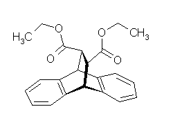
9,10-Dihydro-9,10-ethanoanthracene-11,12-trans-dicarboxylic acid diethyl ester

321
Name 9,10-Dihydro-9,10-ethanoanthracene-11,12-trans-dicarboxylic acid diethyl ester
Synonyms
Name in Chemical Abstracts 9,10-Ethanoanthracene-11,12-dicarboxylic acid, diethyl ester, trans-
CAS No 93368-53-7
EINECS No
Molecular formula C22H22O4
Molecular mass 350.42
SMILES code c1cccc2[C@@H]3[C@H](C(=O)OCC)[C@@H](C(=O)OCC)[C@H](c12)c4ccccc43

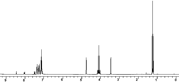
1H NMR

1H NMR

1H-NMR: 9,10-Dihydro-9,10-ethanoanthracene-11,12-trans-dicarboxylic acid diethyl ester
250 MHz, CDCl3
delta [ppm] mult. atoms assignment
1.23 t (3J = 7.2 Hz) 6 H CH3 ethyl
3.45 m 2 H 11-H, 12-H (-CH-COO-)
4.08 m 4 H CH2 ethyl
4.75 m 2 H 9-H, 10-H


13C-NMR

13C NMR

13C-NMR: 9,10-Dihydro-9,10-ethanoanthracene-11,12-trans-dicarboxylic acid diethyl ester
62.5 MHz, CDCl3
delta [ppm] assignment
14.2 CH3 (ethyl ester)
46.7 C9, C10 (CH)
47.7 C11, C12 (CH-COO)
60.9 CH2 (ethyl ester)
123.8 CH arom.
124.5 CH arom.
126.2 CH arom.
126.3 CH arom.
140.3 C quart. arom.
142.0 C quart. arom.
172.3 C(=O)O-
76.5-77.5 CDCl3


IR

IR

IR: 9,10-Dihydro-9,10-ethanoanthracene-11,12-trans-dicarboxylic acid diethyl ester
[KBr, T%, cm-1]
[cm-1] assignment
3074, 3026 arom. C-H valence
2981 aliph. C-H valence
2935, 2897 aliph. C-H valence
1739 C=O valence, ester
1467 arom. C=C valence


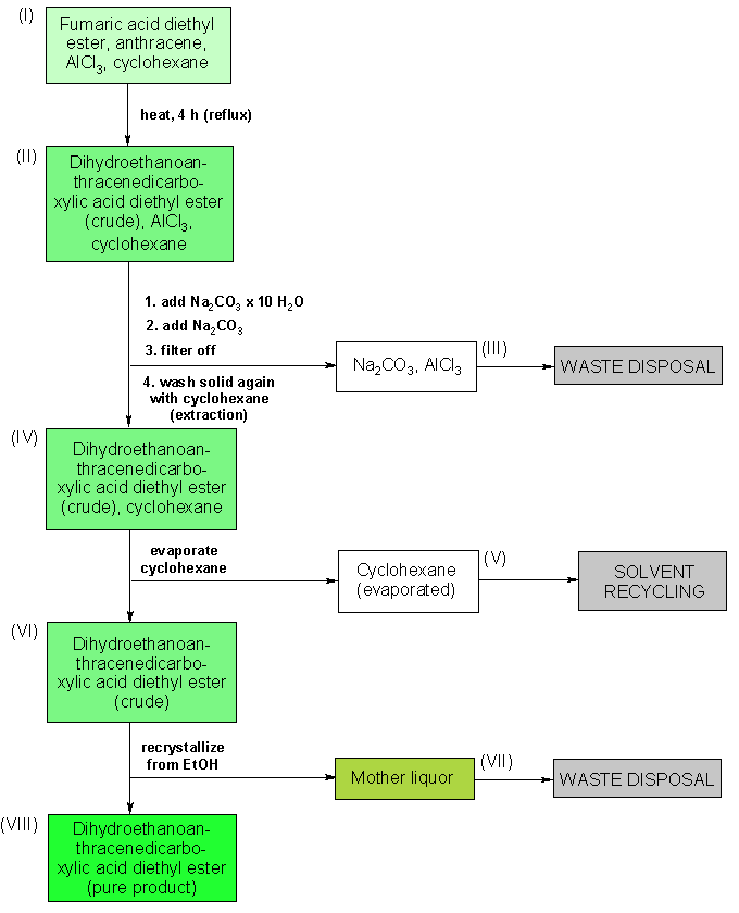
Anthracene + Fumaric acid diethyl ester
reacts to
9,10-Dihydro-9,10-ethanoanthracene-11,12-trans-dicarboxylic acid diethyl ester ; Side reactions


Synthesis of 9,10-dihydro-9,10-ethanoanthracene-11,12-trans-dicarboxylic acid diethyl ester

Reaction type: cycloaddition, Diels-Alder reaction
Substance classes: alkene, aromatics, carboxylic acid ester, diene, dienophile, acid catalyst
Techniques: working with moisture exclusion, heating under reflux, stirring with magnetic stir bar, filtering, evaporating with rotary evaporator, recrystallizing, use of an ice cooling bath, heating with oil bath


Equipment

Batch scale: 0.01 mol 0.1 mol Anthracene
three-necked flask 1000 mL three-necked flask 1000 mL adapter with ground-glass joint and hose coupling adapter with ground-glass joint and hose coupling
protective gas piping protective gas piping reflux condenser reflux condenser
drying tube drying tube bubble counter bubble counter
powder funnel powder funnel heatable magnetic stirrer with magnetic stir bar heatable magnetic stirrer with magnetic stir bar
rotary evaporator rotary evaporator ice bath ice bath
exsiccator with drying agent exsiccator with drying agent oil bath oil bath


Operating scheme

Operating scheme


MULTAN, PAKISTAN


The multan city of saints

Mutlan is an important city of Pakistan which is also known as the city of Saints. The history of Multan begins with the Alexander and later on Kushans, Arabs, Huns, Ghaznavi, Afghans, Mongols, Sikhs, Mughals and British ruled over the city. It is the city of Sufis and Saints who preached the Islam in this region. In the South Asia Multan is the oldest city.

.

Map of Multan

Multan Things to Do

  • Baha-ud-din Zakariya Mazar, Multan

    Baha-ud-din Zakariya Mazar

    Sheikh Baha-ud-din Zakariya (1170-1267) was a sufi saint who for several years travelled the region from Baghdad, Iraq to India preaching Islam, and made his final abode in Multan. His offsprings and disciples travelled all over India to preach. His most famous descendant is Shah…  more

  • Shah Rukn-e-Alam's tomb, Multan

    Shah Rukn-e-Alam’s tomb

    Situated on top of a small hillock, behind the old ruins of Multan Fort, the Mazar and its majestic dome is the first landmark visible when you enter proper Multan. Shah Rukn-e-Alam (1251-1335) was a Sufi saint in Multan. He was revered by his followers, and to this date thousands of pilgrims from all over…  more

  • Other Saints and Sufis Mazars, Multan

    Other Saints and Sufis Mazars

    Multan is known as City of Saints, and this is evident by the number of Masuleums, Mazars, Dargas, or tombs situated in this city. Some of them are listed below.1 Hazrat Baha-ud-Din Zakaria2 Shah Rukn-i-Alam3 Shah Shams Sabzwari4 Shah Gardez5 Musa Pak Shaheed6 Hazrat Hafiz Muhammad…  more

  • Ghanta Ghar (Clock Tower house), Multan

    Ghanta Ghar (Clock Tower house)

    Ghanta Ghar whch is situated in the city center is the city government head quarter. (not a great picture, taken from a moving car)

     more

  • Multan Fort, Multan

    Multan Fort

    The Multan Fort on a high mound of earth which separated it from the old branch of the river Ravi. There are now only remnant of this old fort, which was considered as one of the best fort (defense wise) built in the sub-continent. The fort was destroyed when the British took over. During its haydays the fort walls were was almost 1.6…  more

  • King mosque Eid Gah, Multan

    King mosque Eid Gah

    This Masjid is a marvelous piece of architecture of multan.it is a very beautiful masjid and must to visit place of Multran city

     more

  • Shrines must to visit, Multan

    Shrines must to visit

    Given below is list of must to visit shrines in Multan1.Shrine B.B Pak Damman2.Shrine Hameed-ud-Din Hakim3.Shrine Qutab-al-quteeb’Moj Daryan’4.Shrine-Syed Pir Sakhi Shah Hasan Prwana5.Shrine-Qazi Qutab-ud-Din Kashani6.Shrine-Syed Hasan Kanjzee7.Shrine-Hazarat Shah Dana Shaheed8.Shrine-Abu…  more

  • Shrines of the sufi hermits, Multan

    Shrines of the sufi hermits

    Hi Awais,All the destinations in Pakistan, they’ve their own attraction or somethijng very special in that area, likewise in Hunza, you can’t find the shrines of the sufi hermits, or in Multan, the mountains like Nanga parbat or Kalash tribes in Lahore?Multan, Medina-tul-awlia, the city of saints, famous all over the…  more

  • Visiting Historical Places, Multan

    Visiting Historical Places

    As I describe before that Multan is the city of Saints so there r so many Tombs to Visit and explore the history.

     more

 Multan Hotels

////////


  DRUG APPROVALS BY DR ANTHONY MELVIN CRASTO …..FOR BLOG HOME CLICK HERE


Join me on Linkedin

View Anthony Melvin Crasto Ph.D's profile on LinkedIn

Join me on Facebook FACEBOOK

Join me on twitterFollow amcrasto on Twitter

Join me on google plus Googleplus

 amcrasto@gmail.com

Share

NMR, 3-[3-(benzoylamino)-4-hydroxylphenyl] propanoic acid

 spectroscopy  Comments Off on NMR, 3-[3-(benzoylamino)-4-hydroxylphenyl] propanoic acid
Jul 022015


http://www.google.com/patents/WO2008022462A1?cl=en


1 H-NMR Spectrum of Compound 35…………3-[3-(benzoylamino)-4-hydroxylphenyl] propanoic acid

1H-NMR (Acetone-D6) δ: 2.60 (t, 2H, J = 7.4, H- 3), 2.84 (t, 2H, J = 7.9, H-2), 6.89 (d, IH, J = 8.2, H-8), 7.00 (dd, IH, J = 2.1 , 8.25, H- 9), 7.57 (m, 4H, H-5, H-4′, H-5′, H-61), 8.05 (d, 2H, J = 8.2, H-3′, H-7′), 9.07 (broad s, IH, NH), 9.54 (broad s, IH, OH), 10.58 (broad s, IH, CO2H).

Figure imgf000063_0001
Figure imgf000063_0002

13C-NMR Spectrum of Compound 35

13C-NMR (Acetone- D6) δ: 30.87 (C- 3), 36.21 (C- 2), 118.69 (C- 8), 123.31 (C-5), 123.41 (C- 6), 126.88 (C- 9), 127.37 (C- 4), 128.54 (C-41, C-61), 129.61 (C-31, C-7′), 132.99 (C-51), 134.99 (C-21), 148.03 (C-7), 167.34 (C-I1), 173.94 (C-I ).

Figure imgf000064_0001
Figure imgf000064_0002

13C-NMR Spectrum of Compound 35

Figure imgf000065_0001
Figure imgf000065_0002

COSY-NMR Spectrum of Compound 35

Figure imgf000066_0001
Figure imgf000066_0002

COSY-NMR Spectrum of Compound 35


Figure imgf000067_0001

HETCOR-NMR Spectrum of Compound 35


Figure imgf000068_0001


3-[3-(benzoylamino)-4-hydroxylphenyl] propanoic acid 35:


To a solution of 32 (222 mg, 1.06 mmol, leq.) dissolved in THF (20 mL) was added the catalyst 10 % palladium-on-charcoal (15 % by mass, 33 mg). The resulting mixture was then placed on a hydrogenator, flushed (5 times) with hydrogen and left to agitate under pressure (36 psi.) overnight (12 hrs) while recharging hydrogen pressure twice (36 psi.) until hydrogen up-take by reaction mixture stopped (pressure did not decrease for 1-2 hrs.). The reaction mixture was vacuum filtered through Celite ‘ rinsing with THF. To the filtered solution containing 33 was directly added BzCl (154 mg, 1.1 mmol, 1 eq.) and left to stir at room temperature for 30 min. Then 10 % HCl (25 mL) was added and stirring continued an additional 5 min. followed by extraction with CIT2Cl2 (2 x 35 mL). The organic fractions were combined, dried (MgSO4), and evaporated off solvent. The resulting mixture was re-crystallized with Hexane/ Acetone to afford an off white solid (250 mg) with an 83 % yield from compound 32. Molecular Formula – C16Hi5NO4. Formula Weight – 285.295 g mole“1.

FT-IR (KBR disk) cm” 1 : 3201 (NH, OH), 1692 (CO2H), 1636 (NHAc).

1H-NMR (Acetone-D6) δ: 2.60 (t, 2H, J = 7.4, H- 3), 2.84 (t, 2H, J = 7.9, H-2), 6.89 (d, IH, J = 8.2, H-8), 7.00 (dd, IH, J = 2.1 , 8.25, H- 9), 7.57 (m, 4H, H-5, H-4′, H-5′, H-61), 8.05 (d, 2H, J = 8.2, H-3′, H-7′), 9.07 (broad s, IH, NH), 9.54 (broad s, IH, OH), 10.58 (broad s, IH, CO2H).

13C-NMR (Acetone- D6) δ: 30.87 (C- 3), 36.21 (C- 2), 118.69 (C- 8), 123.31 (C-5), 123.41 (C- 6), 126.88 (C- 9), 127.37 (C- 4), 128.54 (C-41, C-61), 129.61 (C-31, C-7′), 132.99 (C-51), 134.99 (C-21), 148.03 (C-7), 167.34 (C-I1), 173.94 (C-I ).

/////////////////////////////////////////


1H-NMR Spectrum of Compound (+/-V36

Figure imgf000069_0001
Figure imgf000069_0002

13 C-NMR Spectrum of Compound (+/-V36

Figure imgf000070_0001
Figure imgf000070_0002

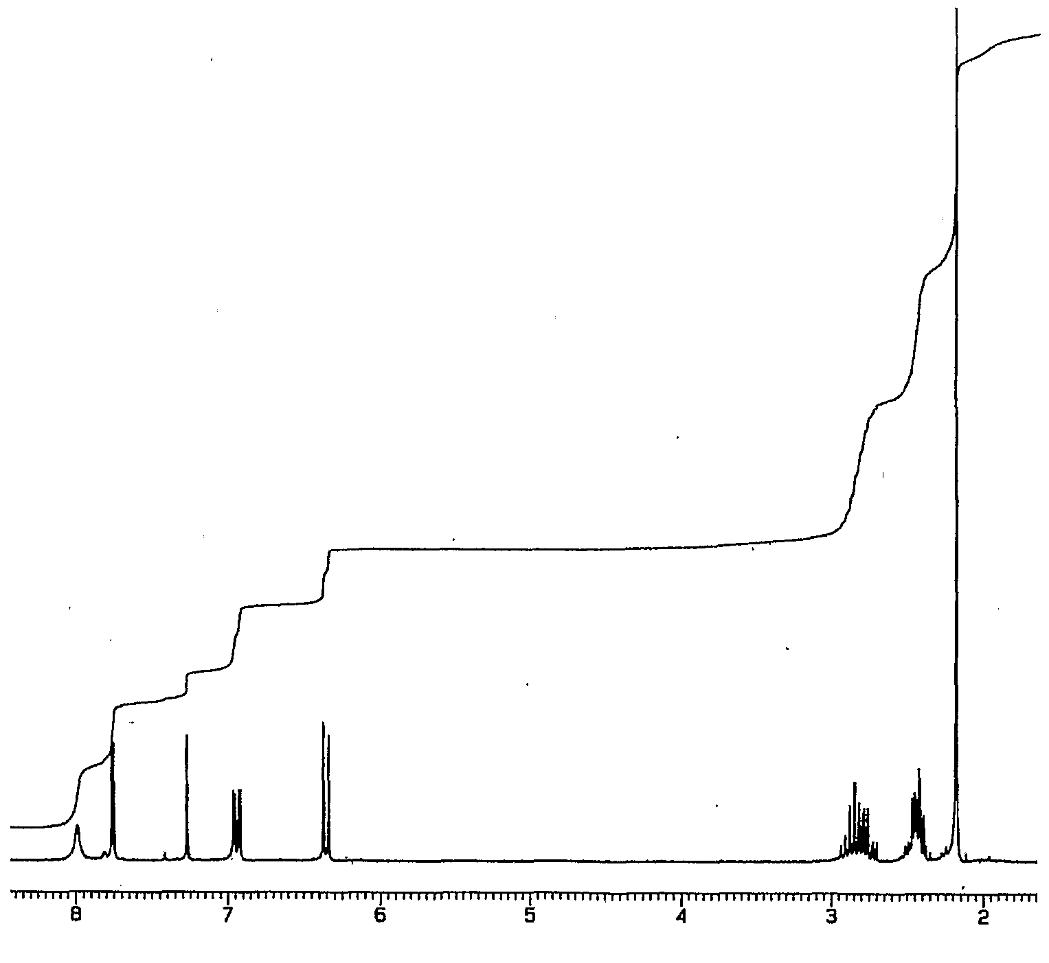
N-(l-oxaspiro[4.5]deca-6,9-dien-2,8-dion-7-yl)acetamide (+/-)-36: To a solution of 34 (122 mg, .547 mmol, 1 eq.) dissolved in acetone (10 mL, 0 0C) was added PIFA (306 mg, .71 1 mmol, 1.3 eq.) in one portion and stirred for 20-25 minutes (confirmed by tic: [1 : 1] EtOAc/Hexane). The reaction mixture was diluted with ethyl acetate (15 mL), washed with cold water (10 mL), dried organic fraction (MgSO4) and evaporated off solvent to afford a Tan solid. The crude product was purified by re-dissolving with CHCI3, filtering of the solution through Celite ®, evaporating off the solvent and placing it under vacuum overnight to afford an off white solid (120 mg, 98 % yield). Molecular Formula – C1 1Hi iNO4. Formula Weight – 221.209 g mole“1. FT-IR (KBR disk) cm“1: 3333 (NH), 1777 (lactone), 1668 (amide), 1650 (ketone), 1620 (α, β-conjugation to ketone). 1H-NMR (CDCl3) δ: 2.17 (s, 3H, H-2′), 2.44 (m, 2H, H-4), 2.81 (m, 2H, H-3), 6.35 (d, IH, J = 10.0, H-9), 6.94 (dd, IH, J = 3.1, 10.0, H- 10), 7.75 (d, IFI, J = 3.1, H-6), 7.99 (broad s, IH, NH). 13C-NMR (CDCl3) δ: 24.86 (C- 2′), 28.36 (C- 4), 32.91 (C- 3), 79.76 (C-5), 124.30 (C- 6), 127.12 (C- 9), 131.55 (C- 7), 148.37 (C-10), 169.51 (C-I’), 175.46 (C-2), 179.40 (C- 8).


////////////////////

1H-NMR Spectrum of Compound 32

Figure imgf000059_0001
Figure imgf000059_0002

(2E)-3-(4-hydroxyl-3-nitrophenyl) acrylic acid 32: To a solution of 4- hydroxyl-3-nitrobenzaldehyde (1.073 g, 6.43 mmol, 1 eq.) dissolved in pyridine (25 mL) was added piperidine (25 drops) and the resulting mixture was stirred (4-5 min.). Malonic acid (1.671 g, 16.1 mmol, 2.5 eq.) was then added in one portion and the resulting mixture was warmed (60-63 0C) and stirred overnight (12-14 hrs, confirmed by tic: EtOAc, mini work up, 10 % HCl and EtOAc). The reaction was cooled and acidified (50 % HCl) until yellow precipitate formed (pH~2). This yellow precipitate was extracted with ethyl acetate (2 x 150 niL). The organic fractions were combined and washed with brine (150 mL), dried (MgSO4), and the solvent was evaporated to afford a yellow solid. Removed excess solvent by vacuum and used without further purification (1.250 g, 93 % yield). Molecular Formula – CgH7NO5. Formula Weight – 209.156 g mole“1. FT-IR (KBR disk) cm“1: 2942 (OH), 1684 (CO2H), 1626 (C=C), 1533,1270 (NO2). 1FI-NMR (Acetone-D6) δ: 2.87 (broad s, IH, OH), 6.58 (d, IH, J= 16.0, H-2), 7.27 (d, IH, J= 8.8, H-8), 7.70 (d, IH, J= 16.4, H-3), 8.08 (d, IH, J= 2.2, 8.5, H-9), 8.40 (d, IFI, J = 2.2, FI-5), 10.67 (broad s, I H, CO2FI). The13C-NMR of this compound agrees with the previously published data.52


Con Dao Island, Vietnam


con dau six senses resort image

Con Dao Island, Vietnam


This 16-island archipelago is a “pocket of paradise,” says Robert Reid, a travel editor at Lonely Planet.

Getting there: Take a 45-minute flight from Ho Chi Minh City.

What to do: The diving is among the best in Vietnam. Take scuba lessons as a couple or discover the nearby secluded beaches of Bai Dat Doc and Dam Trau.

Where to stay: Six Senses resort offers luxury villas on the East Vietnam Sea. The resort has an in-house spa offering traditional Vietnamese healing practices; it also boasts outdoor treatment rooms and a yoga and meditation pavilion. Inquire for rates.


Con Dao travel guide – Wikitravel

wikitravel.org/en/Con_Dao

Con Dao is an island off the southern coast of Vietnam. … The Con Dao Islands separated from the mainland about 15,000 years ago. This has resulted in the …

Understand – ‎Get in – ‎Get around – ‎See


.


airport


////////////

Share

Nonanedioic acid, Azelaic acid

 spectroscopy, SYNTHESIS  Comments Off on Nonanedioic acid, Azelaic acid
Jun 292015


148


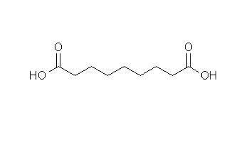
Nonanedioic acid

Azelaic acid

148
Name Nonanedioic acid
Synonyms Azelaic acid
Name in Chemical Abstracts Nonanedioic acid
CAS No 123-99-9
EINECS No 204-669-1
Molecular formula C9H16O4
Molecular mass 188.23
SMILES code O=C(O)CCCCCCCC(=O)O
Ricinolic acid
KMnO4 / KOH
reacts to
Nonanedioic acid ; Side reactions

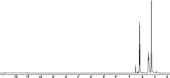
1H NMR

1H NMR

1H-NMR: Nonanedioic acid
250 MHz, DMSO-d6
delta [ppm] mult. atoms assignment
1.25 m 6 H 4-H, 5-H, 6-H
1.47 m 4 H 3-H, 7-H
2.18 t (3J = 7.3 Hz) 4 H 2-H, 8-H
ca. 12 broad s 2 H COOH
2.49 DMSO


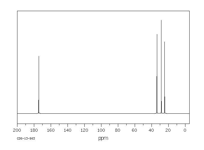
13C-NMR

13C NMR

13C-NMR: Nonanedioic acid
62.5 MHz, DMSO-d6
delta [ppm] assignment
23.9 C3, C7
27.6 C5
27.8 C4, C6
33.1 C2, C8
173.4 COOH
39.5 DMSO-d6

Azelaic acid(123-99-9)13CNMR

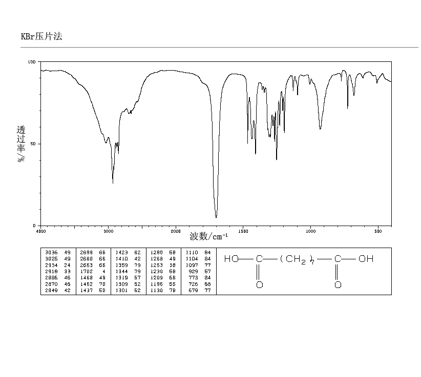
IR

IR

IR: Nonanedioic acid
[KBr, T%, cm-1]
[cm-1] assignment
3300-2500 O-H valence, superimposed on C-H valence
2962, 2887 aliph. C-H valence
1724 C=O valence, carboxylic acid

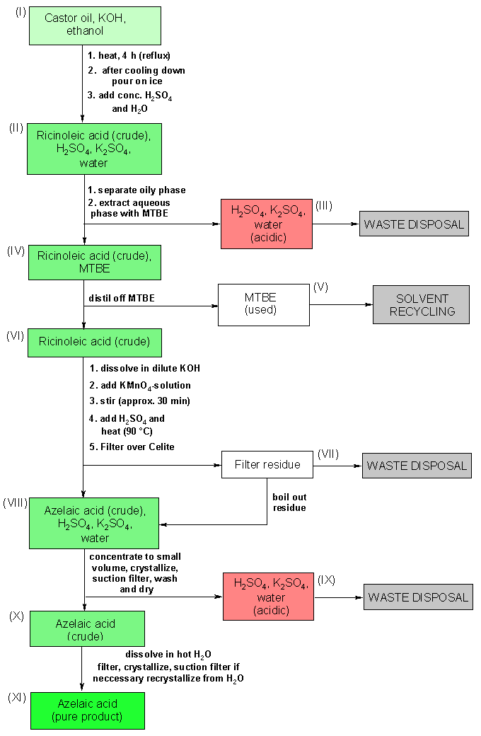
Oxidation of ricinoleic acid (from castor oil) with KMnO4 to azelaic acid

Reaction type: oxidation
Substance classes: alkene, carboxylic acid, renewable resources
Techniques: heating under reflux, stirring with magnetic stir bar, stirring with KPG stirrer, adding dropwise with an addition funnel, shaking out, extracting, evaporating with rotary evaporator, filtering, recrystallizing, heating with oil bath
Degree of difficulty: Medium


Operating scheme

Operating scheme


Equipment

Batch scale: 0.04 mol Ricinolic acid
round bottom flask 250 mL round bottom flask 250 mL three-necked flask 1000 mL three-necked flask 1000 mL
reflux condenser reflux condenser internal thermometer internal thermometer
addition funnel with pressure balance addition funnel with pressure balance heatable magnetic stirrer with magnetic stir bar heatable magnetic stirrer with magnetic stir bar
KPG stirrer KPG stirrer beaker 400 mL beaker 400 mL
beaker 250 mL beaker 250 mL Erlenmeyer flask 250 mL Erlenmeyer flask 250 mL
separating funnel separating funnel rotary evaporator rotary evaporator
suction filter suction filter suction flask suction flask
exsiccator with drying agent exsiccator with drying agent oil bath oil bath

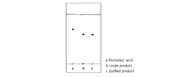
Chromatogram

crude product chromatogram

TLC: crude product
TLC layer Merck silica gel 60 F254, 5 x 10 cm
mobile phase EtOH
staining reagent 0.1% solution of 2,6-dichlorophenolindophenol sodium salt in 95% ethanol
Rf (educt) 0.70
Rf (product) 0.60

PESHAWAR, PAKISTAN FOOD


Peshawar is one the oldest cities of South Asia. It is an entrance point of Pakistan from the Afghanistan. It was an important city of Subcontinent and a meeting and marketing place for the public of Middle East, India and central Asia. Afghan warriors used this way to enter into subcontinent.

.

NALLI GOSHT

Pakistani cooks prepare food for refugees in the Jalozai camp in Peshawar,

Food being prepared at Qissa Khuwani Bazaar in Peshawar on the eve of Eid Milad-

The Big Pot Tea Man of Peshawar

//////

  DRUG APPROVALS BY DR ANTHONY MELVIN CRASTO …..FOR BLOG HOME CLICK HERE


Join me on Linkedin

View Anthony Melvin Crasto Ph.D's profile on LinkedIn

Join me on Facebook FACEBOOK

Join me on twitterFollow amcrasto on Twitter

Join me on google plus Googleplus

 amcrasto@gmail.com


/////////

Share

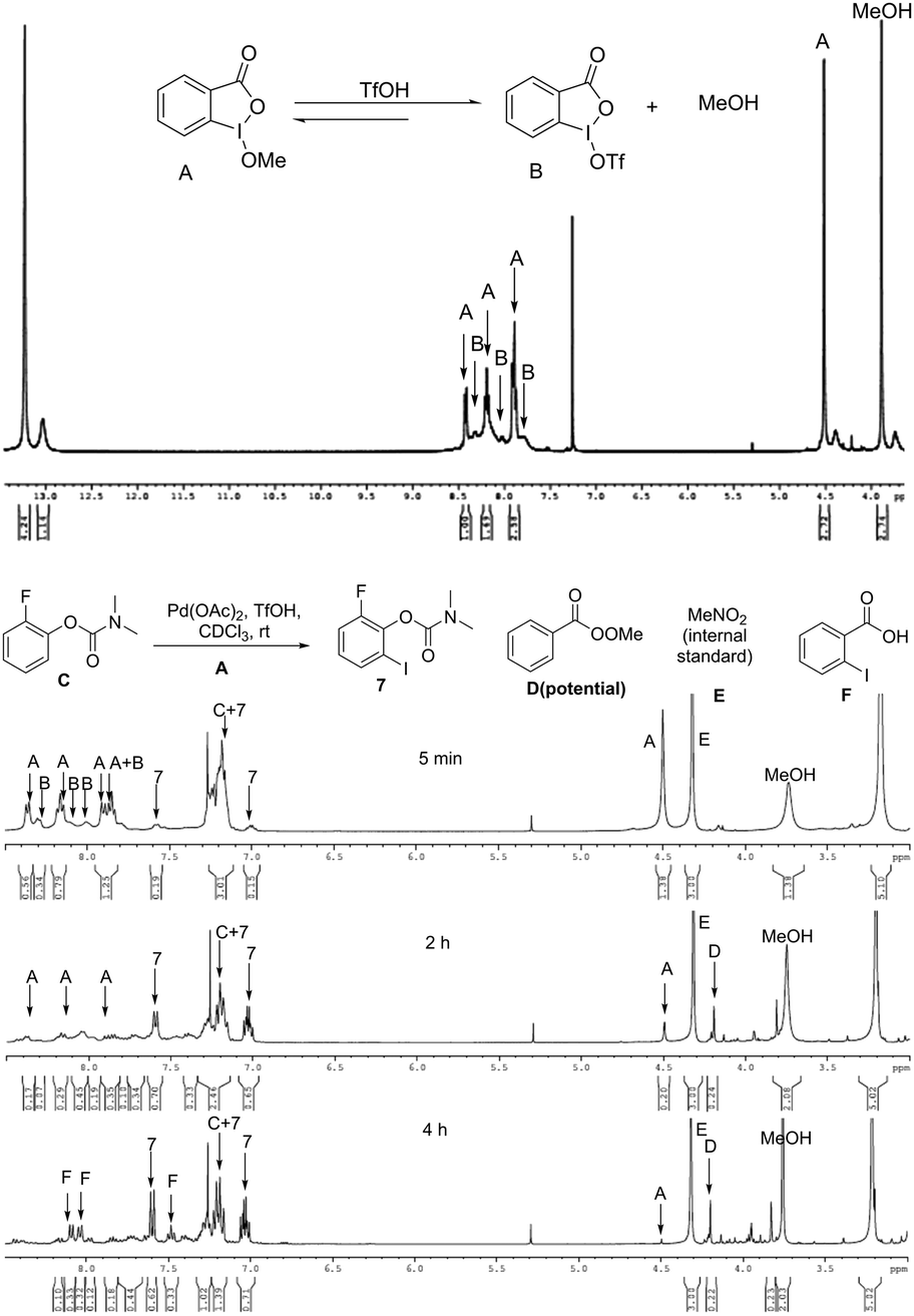
Pd(II) catalyzed ortho C–H iodination of phenylcarbamates at room temperature using cyclic hypervalent iodine reagents

 spectroscopy, SYNTHESIS  Comments Off on Pd(II) catalyzed ortho C–H iodination of phenylcarbamates at room temperature using cyclic hypervalent iodine reagents
Jun 292015
 

A novel approach to access ortho iodinated phenols using cyclic hypervalent iodine reagents through palladium(II) catalyzed C–H activation has been developed through weak coordination. The reaction showed excellent regioselectivity, reactivity and good functional group tolerance. A unique mechanism was proposed.

Graphical abstract: Pd(ii) catalyzed ortho C–H iodination of phenylcarbamates at room temperature using cyclic hypervalent iodine reagents

Pd(II) catalyzed ortho C–H iodination of phenylcarbamates at room temperature using cyclic hypervalent iodine reagents

Xiuyun Sun,a   Xia Yao,a   Chao Zhanga and   Yu Rao*a
*Corresponding authors
aMOE Key Laboratory of Protein Sciences, Department of Pharmacology and Pharmaceutical Sciences, School of Medicine and School of Life Sciences, Tsinghua University, Beijing 100084, China
Chem. Commun., 2015,51, 10014-10017

DOI: 10.1039/C5CC02533H

Rao, Yuyu rao
tsinghua univerisity school of medicines logo

Zhang Chao

 Tsinghua University, Beijing
///
Share

2-Hydroxymalonitrile-A Useful Reagent for One-step Synthesis of α-Hydroxy Esters

 spectroscopy, SYNTHESIS  Comments Off on 2-Hydroxymalonitrile-A Useful Reagent for One-step Synthesis of α-Hydroxy Esters
Jun 042015


YANG Jianxin, YIN Yunxing, HE Zhenmin, MA Li, LI Xin, ZHANG Zhiliu, LIN Xiaojuan, MA Rujian
2-Hydroxymalonitrile-A Useful Reagent for One-step Synthesis of α-Hydroxy Esters
2015 Vol. 31 (3): 321-324 [Abstract] ( 47 ) [HTML 1KB] [PDF 0KB] ( 68 )
doi10.1007/s40242-015-4495-6

see

http://www.cjcu.jlu.edu.cn/hxyj/EN/abstract/abstract16155.shtml


2-Hydroxymalonitrile-A Useful Reagent for One-step Synthesis of α-Hydroxy Esters
YANG Jianxin1,2, YIN Yunxing2, HE Zhenmin2, MA Li2, LI Xin2, ZHANG Zhiliu2, LIN Xiaojuan2, MA Rujian2
1. Tianjin Key Laboratory for Modern Drug Delivery & High-Efficiency, School of Pharmaceutical Science and Technology, Tianjin University, Tianjin 300072, P. R. China;
2. WuXi PharmaTech Co. Ltd., Shanghai 200131, P. R. China
Corresponding Authors: MA Rujian     E-mail: marj@wuxiapptec.com


TAKE A TOUR


Tianjin china

Map of tianjin

Image result for tianjin china map.


.


/////////

Share

KETO ENOL TAUTOMERISM AND NMR

 spectroscopy, Uncategorized  Comments Off on KETO ENOL TAUTOMERISM AND NMR
Jun 032015
 

.


H Nmr Spectrum | Apk Mod Game

www.apkmodgame.net

Shows a method for getting all the useful information out of a proton nmr spectrum and using it to piece together the identity of an unknown molecule.
A Partial NMR Spectrum of 2,4-Pentanedione


Patent EP0922715B1 – Stimuli-responsive polymer utilizing keto …

Carbonyl compounds (aldehydes, ketones, carboxylic esters, carboxylic amides) react aselectrophiles at the sp2 hybridized carbon atoms and as nucleophiles if they contain an H-atom in the α-position relative to their C=O or C=N bonds. This is because this H is acidic and it can be removed by a base leaving behind an electron pair for nucleophilic attacks.

For most compounds in organic chemistry all the molecules have the same structure – even if this structure cannot satisfactory represented by a Lewis formula – but for many compounds there is a mixture of two or more structurally distinct compounds that are in rapid equilibrium. This phenomenon is called tautomerism.

Tautomerism is the phenomenon that occurs in any reaction that simply involves the intramolecular transfer of a proton. An equilibrium is established between the two tautomers (structurally distinct compounds) and there is a rapid shift back and forth between the distinct compounds.

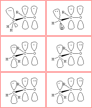
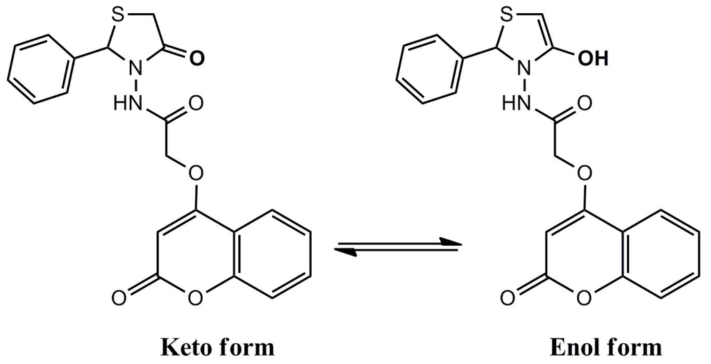
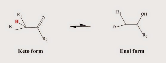
A very common form of tautomerism is that between a carbonyl compound containing an αhydrogen and its enol form (Fig. I.1).

Fig. I.1: A keto-enol reaction


An enol is exactly what the name implies: an ene-ol. It has a C=C double bond (diene) and an OH group (alcohol) joined directly to it.

Notice that in the above reaction as in any keto-enol reaction there is no change in pH since a proton is lost from carbon and gained on oxygen. The reaction is known as enolization as it is the conversion of a carbonyl compound into its enol.

Notice also that in the above reaction the product is almost the same as the starting material since the only change is the transfer of one proton and the shift of the double bond.

In simple cases (R2 = H, alkyl, OR, etc.) the equilibrium of the keto-enol reaction lies well to the left (keto structure) (Table I.1). The reason can be seen by examining the bond energies in Table I.2.


Compound

Enol Content, %

Acetone

6 * 10-7

PhCOCH3

1.1 * 10-6

CH3CHO

6 * 10-5

Cyclohexanone

4 * 10-5

Ph2CHCHO

9.1

PhCOCH2COCH3

89.2

Table I.1: The enol content of some carbonyl compounds


If keto-enol reactions are common for aldehydes and ketones why don’t simple aldehydes and ketones exist as enols?

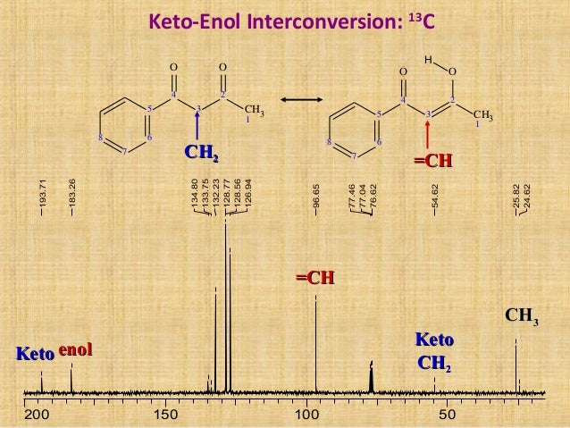
IR and NMR Spectra of carbonyl compounds show no signs of enols. The equilibrium lies well over towards the keto form (the equilibrium constant k for cyclohexanone is about 10-5).


Bond (Energy, kJ/mol)

Sum ( kJ/mol)

keto form

C-H (413)

C-C (350)

C=O (740)

1503

enol form

C=C (620)

C-O (367)

O-H (462)

1449

Table I.2: Bond energies in the keto and in the enol form. The keto form is thermodynamically more stable than the enol form by approximately 50 kJ/mol

The approximate sum of the bond energies in the keto form is 1503 kJ/mol while in the enol form 1449. Therefore, the keto form is thermodynamically more stable than the enol form by approximately 50 kJ/mol.

In most cases, enol forms cannot be isolated since they are less stable and are formed in minute quantities. However, there are some exceptions and in certain cases a larger amount of the enol form is present and it can be even the predominant species:

  • Molecules in which the enolic double bond is in conjugation with another double bond (cases are shown in Table I.1 like Ph2CHCHO and PhCOCH2COCH3)
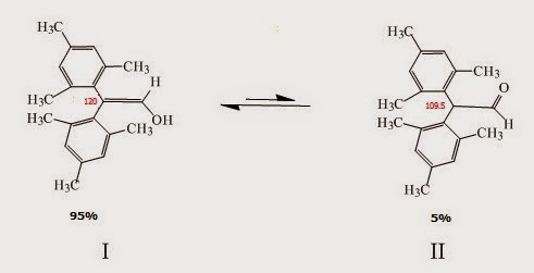
  • Molecules that contain two or more bulky aryl groups (Fig. I.2). Compound I in Fig. I.2 (a substituted enol) is the major species in equilibrium (~95%) while the keto form is the minor species (~5%). In cases like this steric hindrance destabilizes the keto form (the two substituted aryl groups are 109° apart) while in the enol form 120° apart.


Fig. I.2: A keto-enol reaction. The enol form (I) is the major species since the keto form is destabilized by steric hindrance (the substituted aryl groups are closer in the keto form – the C-C angle is 109° and this is unfavorable due to steric hindrance)
Fig. I.2: A keto-enol reaction. The enol form (I) is the major species in this case since the keto form is destabilized by steric hindrance (the substituted aryl groups are closer in the keto form – the C-C angle is 109° and this is unfavorable due to steric hindrance)


Is there experimental evidence that keto-enol reactions are common for aldehydes and ketones?

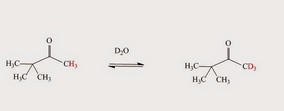
If the NMR spectrum of a simple carbonyl compound in D2O is obtained – such as pinacolone’s (CH3)3CCOCH3 – the signal for protons next to the carbonyl group very slowly disappears. The isolated compound’s mass spectrum (after the above mentioned reaction with D2O is over) shows that those hydrogen atoms have been replaced by deuterium atoms. There is a peak at (M+1)+ or (M+2)+ or (M+3)+ instead of M+. The reaction is shown in Fig. I.3:

 Fig. I.3: Evidence for a keto-enol reaction when pinacolone (CH3)3CCOCH3 reacts with D2O. When the enol form of the pinacolone reverts to the keto form it picks up a deuteron instead of a proton because the solution consists almost entirely of D2O.
Fig. I.3: Evidence for a keto-enol reaction when pinacolone (CH3)3CCOCH3 reacts with D2O. When the enol form of the pinacolone reverts to the keto form it picks up a deuteron instead of a proton because the solution consists almost entirely of D2O.


What mechanism can be proposed for the above reaction?

Enolization is a slow process in neutral solution, even in D2O, and is catalyzed by acid or base in order to happen.

In the acid-catalyzed reaction the molecule is first protonated on oxygen and then loses the C-H proton in a second step (Fig. I.4). When the enol form reverts to the keto – since this is an equilibrium process – it picks up a deuteron instead of a proton since the solution is D2O.


Fig. I.4: The acid-catalyzed keto-enol reaction mechanism. If D2O is the solvent then the α-hydrogens to carbonyl group are replaced by deuterium.
Fig. I.4: The acid-catalyzed keto-enol reaction mechanism. If D2O is the solvent then the α-hydrogens to carbonyl group are replaced by deuterium.

In the base-catalyzed reaction the C-H proton is removed first by the base (for example hydroxide ion OH, OD in our case) and the proton (or D+ in our case) added to the oxygen atom in a second step (Fig. I.5).

Fig. I.5: The base-catalyzed keto-enol reaction mechanism. If D2O is the solvent then the α-hydrogens to carbonyl group are replaced by deuterium.
Fig. I.5: The base-catalyzed keto-enol reaction mechanism. If D2O is the solvent then the α-hydrogens to carbonyl group are replaced by deuterium.

Notice that the enolization reactions in Fig. I.4 and Fig. I.5 are catalytic. In the acid-catalyzed mechanism the D+ (or H+ if water is the solvent) is regenerated at the end (catalyst). In the base-catalyzed mechanism OD (or OH if water is the solvent) is regenerated at the end (catalyst).

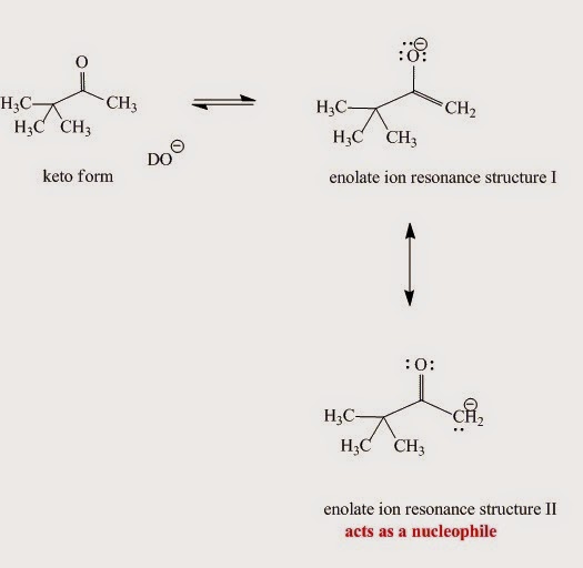
The enolate ion generated from the enol (Fig. I.6) in the base-catalyzed mechanism is nucleophilic due to:

  • Oxygen’s small atomic radius
  • Formal negative charge

An enolate ion is an ion with a negative charge on oxygen with adjacent C-C double bond.


 Fig. I.6: Enolate ion resonance contributors. Although the major contributor is resonace structure I when it reacts as a nucleophile structure II is more reactive.
Fig. I.6: Enolate ion resonance contributors. Although the major contributor is resonace structure I when it reacts as a nucleophile structure II is more reactive.

Enolates are reactive nucleophiles. Although the major enolate Lewis contributor shows concentration of electron density on the electronegative oxygen when it reacts as a nucleophile, it behaves like the electron density is concentrated on the α-carbon next to carbonyl group.

Enolates react with alkyl halides, aldehydes/ketones and esters and these reactions are shown in the post entitled “The chemistry of enolate ions – Enolate ion reactions”.


References
  1. A.J. Kresge, Pure Appl. Chem., 63, 213 (1991)
  2. B. Capon, The Chemistry of Enols, Wiley, NY, 307–322 (1990)
  3. S.E. Biali et al., J. Am. Chem. Soc. 107, 1007 (1985).


.

http://www.slideshare.net/chemsant/nmr-dynamic


http://article.sapub.org/10.5923.j.ajoc.20140401.01.html

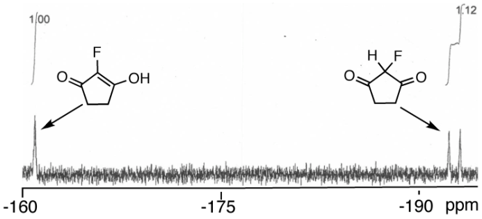
2-fluoro-3-hydroxycyclopent-2-enone and 2-fluoro- 1,3-cyclopentanedione (1c): This compound was obtained as a 52:48 mixture of keto-enol and diketo tautomers in 50% yield as a yellow-brown solid, mp 70-72°C. NMR:1H: δ 2.36 (t, 3JH-H = 16.2 Hz, 2H), 2.85 (m, 2H), 5.91 (d, 2JH-F = 47.7 Hz, 1H). 13C: δ31.1, 90.8 (d, 1JC-F = 251.3 Hz), 122.3 (d, 1JC-F = 233.9 Hz), 210.1 (d, 2JC-F = 31.0 Hz). 19F: keto-enol: δ-161.4 (s, 1F); diketo: δ-195.5 (d, 2JF-H = 47.7 Hz, 1F). Analysis calcd for C5H5FO2: C, 51.73, H, 4.34. Found: C, 51.48, H, 4.31.


Take a tour

Surat gujarat india


Map of surat city.

.

ISKCON

Surat-European tombs


Kathiyavadi food, Garden Restaurant, Restaurant in surat, Restaurant, Restaurant Services, Food


/////////

Share

NMR Structural Techniques’ Contribution To The Drug Discovery And Development Process

 spectroscopy  Comments Off on NMR Structural Techniques’ Contribution To The Drug Discovery And Development Process
Jun 012015
 

Carla Marchioro

Introduction

As is well known the drug discovery and development process is a complex process typically starting from the target identification and validation of a target to progress to clinical studies and hopefully ending with a new drug to the market [Figure 1].


In this process, different approaches and methods are required to understand the disease’s mechanisms, to profile hit molecules that will be progressed to leads suitable for full scale lead optimization programmes and then to generate quality drug candidates to advance to clinical studies.

Focusing on small molecules’ drugs and on hit to candidate phases, a variety of techniques will be used to study the compounds’ profiles at different levels including the physicochemical profiles, purity, solid state behaviours and structures to ensure a quality hit/lead/candidate and related data to allow understanding of the mechanism of action and SAR correlation.

Nuclear Magnetic Resonance (NMR) spectroscopy will play a pivotal role generating data on the molecular interactions between ligands and biological targets, in addition to providing the structures of drug molecules, by-products, impurities, metabolites and quantification data.

NMR Screening Impact 

During the drug discovery phase, NMR spectroscopy is becoming more and more relevant with application at multiple stages along the progression of a project: NMR experiments are used for hits generation, lead discovery and optimization, evaluation of in vitro/in vivo selectivity and efficacy, studies drug toxicity profiles and identification of new drug discovery targets.

Over the last years there has been a large increase in the application of NMR techniques for the rapid determination of protein-ligand structures and interactions, to powerfully screen fragment-based libraries, to identify biological relevant ligand interactions, and to monitor changes in the metabolome from bio-fluids and cells to explore compounds activity.

Focusing on the NMR-based screening techniques, the NMR experiments could be divided into two main categories: target observed and ligand observed methods.

Without doubt, high resolution protein structure is a key requirement to evaluate the biological relevance of a hit from screening and HTS (high-throughput screening) and NMR together with X-ray are playing an essential role.
The last period has witnessed the generation of fast NMR sequences and methods to allow a faster impact on the drug discovery project time but the NMR target-observed techniques still require time, material, possible labelling and difficulties in handling a number of different hits and studies on mixtures.

Nevertheless, if the resonance assignment of the labelled target is known, the exploitation of differences in chemical shifts between free and bound target in two dimensional correlation spectra (shift mapping) will provide important structural information on the site of binding. The experiments could be also be of high value on selectively labelled target decreasing the spectra complexity and so increasing the size of the target that could be studied by NMR techniques.
Considering now that the chemical shift is highly sensitive to the environment of the atom and, as a consequence, it provides information on the binding of a small molecule to a biological target, and on which part of the molecule is interacting and where, it is clear that ligand-observed techniques could generate proof and data for the binding understanding and profile.

In addition, other experiments based on molecule relaxation values are sensitive to the motion of the compound (free vs bound state) and together these experiments will allow validation of ligand binding and/or identification of ligands also in mixtures.

The ligand observed techniques benefit of:

  • one-dimensional experiments;
  • detection of the ligand’s signals (facilitating also the mixture analysis);
  • smaller amount of target substrate;
  • structural and binding information of the ligand;
  • detection of week binding ligands;
  • limited restrictions on size and type of the target, with no isotope labelling requirement or target information details.

On the undesirable side, the techniques could generate false negatives (strong binding and slow exchange equilibria) or false positives (unspecific binding) but all these aspects could be further studied to result in a substantiated answer.

During the Hits generation phase, NMR will be used for the determination of binding affinity values toward the hit validation step to generate a lead where the NMR experiments will also remove the false positives and locate the binding site (for example within the FBDD approach). In the lead optimization phase, to improve potency of the compounds, the epitope mapping will be determined, together with the conformation of the bound ligand, while in the late lead optimization stage for the candidate selection the NMR will support the bioavailability, ADME, PK and toxicological experiments.

The combination of NMR screening methods with other techniques, such as in silico computational protocol, X-ray crystallography, and biophysical experiments will decrease the number of compounds to be studied generating filters and resulting in time and cost saving and efficiency increase. The NMR will be so used to screen and profile a library (or set) of compounds with the unique ability of providing proof for binding between the ligand and the biological target and subsequently being able to detect the binding site and determining the construct of the complex.

This short note will not include technical details of the many NMR-based methods that could be found in several papers and reviews across the last decade [1-8].

The versatility of the NMR techniques is allowing the detection of target-ligand interactions through a large variety of measurements. The insights will derive from the observation of peak intensity and/or line-width changes, saturation transfer differences (STD), chemical shifts perturbations, R2 relaxation effects, R1, sel competition data, induced transferred NOEs, interligand NOEs, diffusion coefficient measurements and changes, and, in general, from monitoring any changes in the NMR spectra resulting from the ligand-target interactions.

A big impact of the NMR techniques is also evident on the “undruggable” targets when other techniques alone fail to result in relevant data and studies on protein-protein, protein-membrane macromolecular recognition are now becoming more and more frequently successfully progressed [9].

The lead compound will need then to be optimized in the bioavailability, efficacy and toxicity profile to result in a candidate to be progressed to in vivo studies, in animals, and finally on humans.

NMR will contribute heavily in all these phases will full characterization of the compound and solid state data, stability studies, formulation studies and NMR-based metabolomics experiments. All these aspects will be covered in a future contribution.

References 

1. M. Pellecchia, I. Bertini, D. Cowburn, C. Dalvit, E. Giralt, W. Jahnke, T.L. James, S.W. Homans, H. Kessler and C. Luchinat, Nat. Rev. Drug Discovery, 7, 738 (2008).

2. R. Powers, Expert Opin. Drug Discov., 4(10), 1077 (2009).

3. R. Powers, J. Med. Chem., 57(14), 5860 (2014).

4. M.J. Harner, A.O. Frank and S.W.Fesik, J. Biomol. NMR, 56(2), 65 (2013).

5. C. Dalvit, Prog. Nucl. Magn. Reson. Spectrosc., 51, 243 (2007).

6. M. Mayer and B. Meyer, Angewandte Chemie Int. Edition, 38,1784 (1999).

7. P.J. Hajduk, D.J. Burns, Comb. Chem. High Throughput Screen., 5, 613 (2002).

8. W.Jahnke and D.A. Erlanson (Editors), Fragment-based Approaches in Drug Discovery, Wiley-VCH, 2006.

9. D.M. Dias, I. Van Molle, M.G.J. Baud, C. Galdeano, C.F. G. C. Geraldes and Alessio Ciulli, ACS Med. Chem. Lett., 5 (1), 23 (2014).

In Part 2 of this series, Carla Marchioro continues to offer her insights into the contribution of NMR structural techniques to the drug discovery and development process.


Introduction 

After some insights on the impact of NMR techniques on the initial drug discovery phase [1], NMR techniques applied in the progression of a compound from lead to candidate and to drug are clearly having, together with other techniques, a large impact with full structural determination, full understanding of chemical reactions, studies of molecules’ behaviour in solutions and solid states and stability monitoring with determination of by-products.

NMR Techniques in Lead Optimization and Drug Development 

As soon as a compound has been identified as a lead to be progressed to the candidate phase, several NMR studies will be required to support the chemical effort, and to ensure a quality profile of the selected compound.

Synthesis of different compounds will be progressed to obtain the desired biological profile and structures will be characterized and studied to also support the computational effort, and to monitor and determine the purity for the biological tests.

Several techniques will be used, such a MS, IR, HPLC,…, to results together with the NMR data in a full profile of the studied compound.

Classical mono- and two-dimensional NMR techniques (1H and 13C) will be performed and, if required, experiments on additional nuclei will add further information to the full structural determination. As an example, in Figures 1 and 2, 1H-15N g-HNMQC, 19F-15N g-HNMQC, and 1H-29Si g-HMQC have been used to obtain the full structures characterizations [2, 3].


Figure 1: 1H-15N g-HNMQC and 19F-15N g-HNMQC experiments.

Figure 1: 1H-15N g-HNMQC and 19F-15N g-HNMQC experiments.


Figure 2: 1H-29Si g-HMQC experiment.

Figure 2: 1H-29Si g-HMQC experiment.


The selected compound(s) will be moved to candidate development with scale-up of the synthetic route, and characterization of the resulting material.

NMR will play an important role in reaction monitoring to ensure, with other techniques, a full understanding of the different steps of the chemical steps with identification of by-products and impurities.

Hyphenated HPLC- NMR has been used in the example in Figure 3 for the identification of co‑eluting low‑level impurities in key intermediate; Spectrum A has been acquired after injection of the mother liquors while Spectrum B has been acquired after injection of 100 µL of a solution of key-intermediate. Detailed analysis on the impurity in the mother liquors with a time-slice HPLC-NMR experiment (3 spectra at 10 sec. interval during peak elution) allowed the confirmation that the impurity was in fact a mixture of two co-eluting products. Structures determination has then been obtained after purification using standard NMR experiments [2].


Figure 3: Identification of co-eluting low-level impurities.

Figure 3: Identification of co-eluting low-level impurities.


Critical experiments are also required in the case of UV transparent compounds, which will not be monitored by classical chromatographic techniques as reported in Figure 4 [2].


Figure 4: Reaction monitoring: Continuous-flow HPLC-NMR.

Figure 4: Reaction monitoring: Continuous-flow HPLC-NMR.


The final API will be fully characterized to profile the solid state profile, and to support the formulation studies. In addition to the solution phase NMR, solid-state NMR (ssNMR) will be used together with a variety of techniques to ensure a full understanding of compound behaviour.

An interesting application of solution NMR is reported in Figure 5 where experiments have been progressed for the determination of the critical micelle concentration (CMC) (value of the solute concentration at which half the total solute is present in the free monomeric form). NMR spectroscopy can be an alternative method to measure the CMC value, being the chemical shift concentration-dependent, particularly in the case of solute-solute intermolecular interactions, with typical downfield shifts of 1H NMR resonances on dilution.


Figure 5: Critical Micelle Concentration (CMC) Determination.

Figure 5: Critical Micelle Concentration (CMC) Determination.

In the example, the particularly large shielding for the aromatic protons allowed the assumption that the aromatic rings of the studied molecules that constitute the aggregate are placed in the inner hydrophobic part of the micelle, while the N-acetylpiperazine ring is somehow representing the hydrophilic external surface of the micelle itself. The forces that are involved in the aggregation are then those typical of π-staking. The CMC can then be evaluated plotting the chemical shift variation (Δδ, ppm) versus the reciprocal of the concentration (L/mol). No significant chemical shift variation was observed in the solutions at concentration ≤ 1 mg/mL, while a linear trend was observed in the concentration range 50 ÷ 3 mg/mL. Thus, assumption could be made that the intercepts of these lines on the x axis corresponded to the 1/CMC value. NMR measurements performed at 15 °C, 25 °C, 35 °C and 45 °C allowed the temperature dependence of the CMC to be determined and the thermodynamic parameters of the micellization process to be extrapolated [4].

In Parts 1 and 2, a few examples of the possibilities of the NMR techniques to support the drug discovery and development have been made with a focus on structures determination and characterization. The impact of NMR techniques on in vitro ex vivo in vivo and clinical phases will be covered in Part 3.

References 

1. C. Marchioro, Spectroscopy Solutions , 3 (1), (2015).

2. S. Provera, C. Marchioro, unpublished data.

3. S. Provera, S. Davalli; G. H. Raza; S. Contini; C. Marchioro, Magn. Reson. Chem. , 39, 38 (2001).

4. S. Provera, S. Beato, Z. Cimarosti, L. Turco, A. Casazza, G. Caivano, C. Marchioro, J. Pharm. Biomed. Anal. , 54, 48 (2011).

Carla Marchioro

Scientific Director at R4R & Head of Discovery and Development; Chief Technology & Operations Advisor at AnCoreX

Current
  1. AnCoreX Therapeutics,
  2. R4R
Previous
  1. Aptuit,
  2. GlaxoSmithKline
Education
  1. Università degli Studi di Padova

https://www.linkedin.com/in/carlamarchioro

Carla Marchioro – ResearchGate

www.researchgate.net/profile/Carla_Marchioro

Carla Marchioro is Scientific Director, and Head of the Pharma & Analytical Division at Research for Rent, R4R, Italy where she is now after covering related positions in Aptuit and GlaxoSmithKline R&D where she has been leading multidisciplinary and cross national groups. In addition, she is also Chief Technology & Operations Adviser at AnCoreX Therapeutics.

She is an NMR expert with a chemistry background and has large experience in structural techniques. Over the years, she has developed an extended experience in a large part of the Research & Development process from target identification and progression to NDA filling.

In her group, in addition to classical structural and analytical approaches, state of the art techniques and technologies such as “omics”, computer-assisted drug design, fragments base screening, analytical and preparative SFC, quantitation by NMR, ssNMR methods for cells & tissues and more have been introduced and developed.

In addition to the R4R role, she is a member of a number of Scientific Boards, European and National Research funding bodies; and she has been part of the Scientific Advisory Board of the ProtEra company up to February 2010.She is author of a number of publications and presentations and she is a well-recognized member in the scientific community. She has been a member of the ENC Scientific Board, the chair of the 51” ENC (2010), member of the SMASH Conference Board, and the chair of the SMASH 2013 Conference.

Structural & Analytical expertise in the Drug Discovery, Chemical Development and Pharmaceutical Development Departments (up to transfer to Manufacturing groups). In addition, experience in the drug design and understanding of mechanism of action, metabolic pathways and safety related aspects.

Specialties: full understanding of mechanism of actions; full understanding of chemical and biological pathways; software and hardware design and needs; international experiences crossing countries and cultures.

Experience

Chief Technology & Operations Advisor

AnCoreX Therapeutics

 – Present (1 year 2 months)

Scientific Director & Head of Discovery and Development

R4R

 – Present (2 years 6 months)

Scientific Liaison Director

Aptuit

 –  (1 year 4 months)Verona Area, Italy

Director, Head of Structural & Analytical Scientific Strategy

Aptuit

 –  (2 years 3 months)

Director Analytical Chemistry

Aptuit

 –  (4 months)

Director & Site Head of Verona Analytical Chemistry

GlaxoSmithKline

 –  (1 year 6 months)

Objective of the Verona group was to provide Structural & Analytical expertises to the Verona/Harlow Centre for Excellence in Drug Discovery (Neurosciences CEDD), Chemical Development and Pharmaceutical Development Departments (up to transfer to Manufacturing groups) at the Verona GSK site. In addition, to contribute to the international initiatives of Molecular Drug Discovery (MDR) and Analytical Chemistry.

Director & Site Head of Verona & Zagreb Analytical Chemistry

GSK

 –  (2 years)

Objective of the Verona group was to provide Structural & Analytical expertises to the Verona/Harlow/Zagreb Centre for Excellence in Drug Discovery (Neurosciences CEDD), Chemical Development and Pharmaceutical Development Departments (up to transfer to Manufacturing groups) at the Verona GSK site. In addition, to contribute to the international initiatives of Molecular Drug Discovery (MDR) and Analytical Chemistry.
Objective of the Zagreb group was to provide Structural & Analytical expertises to the Zagreb Centre for Excellence in Drug Discovery (MacrolidesCEDD), and Pharmaceutical Development Departments at the Zagreb GSK site. In addition, to contribute to the international initiatives of Molecular Drug Discovery (MDR) and Analytical Chemistry.

Director

GlaxoWellcome

 –  (1 year)

Honors & Awards

Additional Honors & Awards

Contract Professor, Ferrara University (1999–2003);
Chiar for the SMASH2003 Conference, Verona, Italy
Chair for the 51th Experimental NMR Conference (ENC) (2010, Daytona Beach, US)
Chair for the SMASH2013 Conference, Santiago de Compostela, Spain

Publications

Discovery Process and Pharmacological Characterization of a Novel Dual Orexin 1 and Orexin 2 Receptor Antagonist Useful for Treatment of Sleep Disorders(Link)

Bioorganic & Medicinal Chemistry Letters (2011), 21, 5562

September 1, 2011

A novel, drug-like bis-amido piperidine derivative was identified as a potent dual OX1 and OX2 receptor antagonists, highly effective in a pre-clinical model of sleep.

VERONA,  ITALY

  1. Verona – Wikipedia, the free encyclopedia

    en.wikipedia.org/wiki/Verona

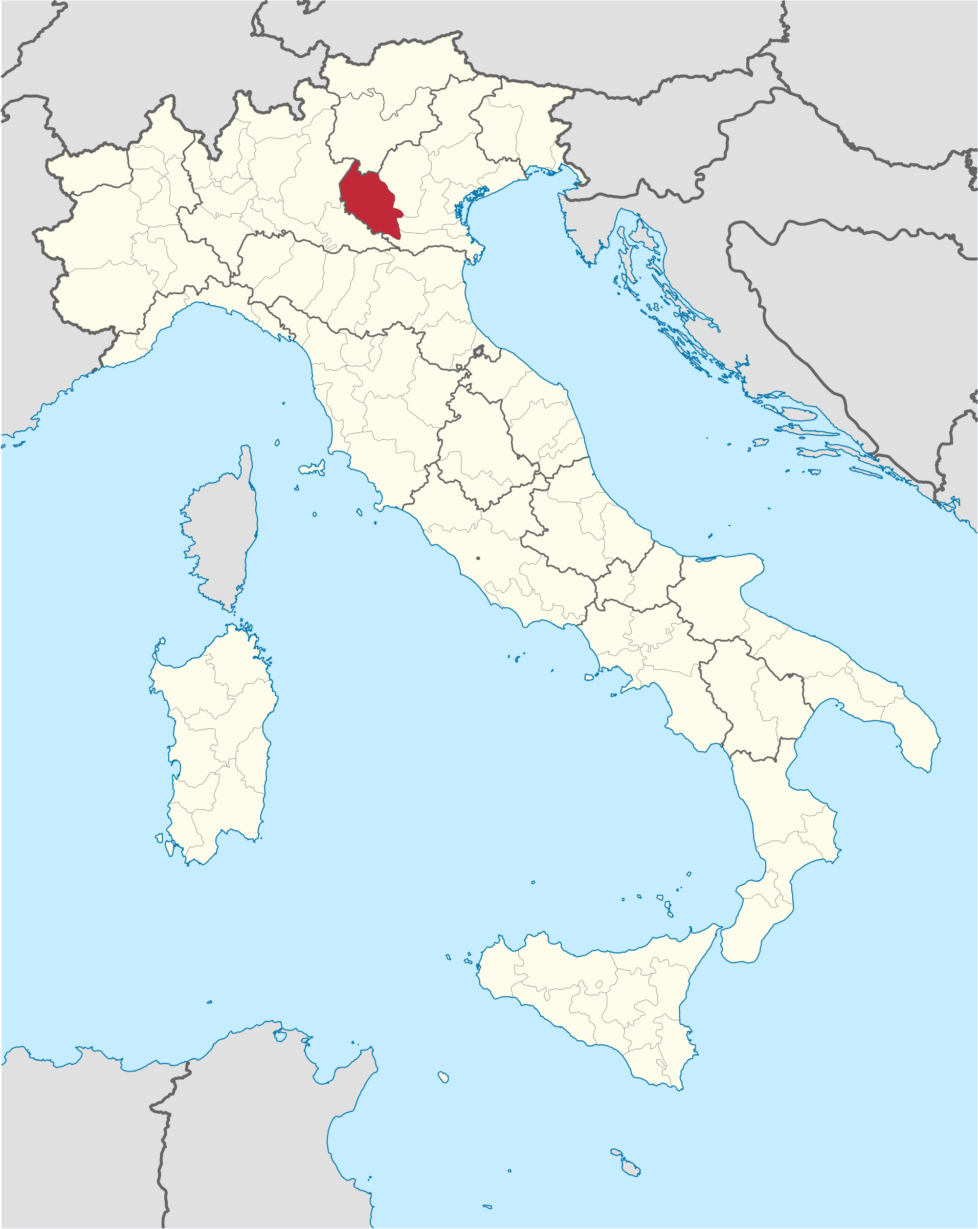
Verona (Italian pronunciation: [veˈroːna] ( listen); Venetian: Verona, Veròna) is a city straddling the Adige river in Veneto, northern Italy, with approximately  …

Map of verona italy.

////////

Share

Simple and effective method for two-step synthesis of 2-(1,3-dithian-2-ylidene)-acetonitrile

 spectroscopy, SYNTHESIS  Comments Off on Simple and effective method for two-step synthesis of 2-(1,3-dithian-2-ylidene)-acetonitrile
Apr 012015
 

Simple and effective method for two-step synthesis of 2-(1,3-dithian-2-ylidene)-acetonitrile

Simple and effective method for two-step synthesis of 2-(1,3-dithian-2-ylidene)-acetonitrile (75% overall yield) and molecular modeling calculation of the mechanism by B3LYP and the 6-311++G(2df,2p) basis set.

http://dx.doi.org/10.5935/0100-4042.20140308

Publicado online: dezembro 12, 2014

Método alternativo para a síntese e mecanismo de 2-(1,3-ditiano-2-ilideno)-acetonitrila

Marcelle S. Ferreira; José D. Figueroa-Villar*
Quim. Nova, Vol. 38, No. 2, 233-236, 2015
Artigo http://dx.doi.org/10.5935/0100-4042.20140308
*e-mail: jdfv2009@gmail.com
MÉTODO ALTERNATIVO PARA A SÍNTESE E MECANISMO DE 2-(1,3-DITIANO-2-ILIDENO)-ACETONITRILA
Marcelle S. Ferreira e José D. Figueroa-Villar* Departamento de Química, Instituto Militar de Engenharia, Praça General Tiburcio 80, 22290-270
Rio de Janeiro – RJ, Brasil
Recebido em 18/08/2014; aceito em 15/10/2014; publicado na web em 12/12/2014
ALTERNATIVE METHOD FOR SYNTHESIS AND MECHANISM OF 2-(1,3-DITHIAN-2-YLIDENE)-ACETONITRILE. We report an alternative method for the synthesis of 2-(1,3-dithian-2-ylidene)-acetonitrile using 3-(4-chlorophenyl)-3-oxopropanenitrile and carbon disulfide as starting materials. The methanolysis of the intermediate 3-(4-chlorophenyl)-2-(1,3-dithian-2-ylidene)-3- oxopropanenitrile occurs via three possible intermediates, leading to the formation of the product at a 75% overall yield. Molecular modeling simulation of the reaction pathway using B3LYP 6-311G++(2df,2p) justified the proposed reaction mechanism. Keywords: 2-(1,3-dithian-2-ylidene)-acetonitrile; reaction mechanism; methanolysis; molecular modeling.
3-(4-clorofenil)-2-(1,3-ditiano-2-ilideno)-3-oxopropanonitrila (3): Cristal amarelo. Rendimento: 95%, 2,80 g, pf 158-160 °C, lit.21 159-160 °C;
IV (KBr, cm-1): 2198 (CN), 1612 (C=O), 1585, 1560 (aromático), 678 cm -1 (C-S);
1H RMN (300 MHz, CDCl3) δ 2,38 (m, J 6,9, 2H, CH2); 3,01 (t, J 6,6, 2H, SCH2); 3,17 (t, J 7,2 , 2H, SCH2); 7,43 (d, J 8,5, 2H); 7,83 (d, J 8,5, 2H);
13C RMN (75 MHz, CDCl3) δ 23,9 (CH2), 30,4 (SCH2), 104,2 (CCO), 117,5 (CN), 128,9, 130,5, 135,6, 139,2 (aromático), 185,2 (C=CS), 185,4 (CO).
21…….Rudorf, W. D.; Augustin, M.; Phosphorus Sulfur Relat. Elem. 1981, 9, 329.
…………………………………….
Síntese da 2-(1,3-ditiano-2-ilideno)-acetonitrila (1) Em um balão de fundo redondo de 100 mL foram adicionados 0,400 g (1,4 mmol) de 3-(4-clorofenil)-2-(1,3-ditiano-2-ilideno)-3- -oxopropanonitrila (2) dissolvidos em 15 mL de THF seco, 0,140 g (20 mmol) de sódio e 15 mL de metanol seco sob atmosfera de nitrogênio. A mistura reacional foi mantida sob agitação à 25 °C por 48 h. Em seguida, a mistura reacional foi dissolvida em 30 mL de água destilada e extraída com acetato de etila (3 x 20 mL). A fase orgânica foi seca em sulfato de sódio anidro, filtrada e concentrada a vácuo para se obter o produto bruto, que foi purificado por cromatografia em coluna (silica gel e hexano:acetato de etila 7:3).
2-(1,3-ditiano-2-ilideno)-acetonitrila (1): Cristal branco. Rendimento: 75%, 165 mg, pf. 60-63 °C, lit1 60-62 °C;
1 H RMN (300 MHz, CDCl3) δ 2,23 (m, J 6,8, 2H, CH2); 3,01 (t, J 7,5, 2H, SCH2); 3,06 (t, J 6,9, 2H, SCH2), 5,39 (s, 1H, CH);
13C RMN (75 MHz, CDCl3) δ 22,9 (CH2), 28,7 (SCH2), 28,8 (SCH2), 90,4 (CHCN), 116,3 (CN), 163,8 (C=CS).
1………Yin, Y.; Zangh, Q.; Liu, Q.; Liu, Y.; Sun, S.; Synth. Commun. 2007, 37, 703.
 Acetonitrile, 1,3-dithian-2-ylidene-

CAS 113998-04-2

  • C6 H7 N S2
  • Acetonitrile, 2-​(1,​3-​dithian-​2-​ylidene)​-
  • 157.26
Melting Point 60-62 °C
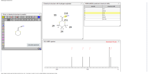
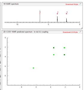
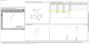
1H  NMR  predict
2-(1,3-dithian-2-ylidene)-acetonitrile
BR 1H
BR 1H 1
ACTUAL 1H NMR VALUES
1 H RMN (300 MHz, CDCl3)
δ 2,23 (m, J 6,8, 2H, CH2);
3,01 (t, J 7,5, 2H, SCH2);
3,06 (t, J 6,9, 2H, SCH2),
5,39 (s, 1H, CH);
……………………..
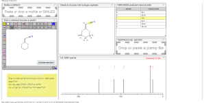
13C NMR PREDICT
BR 13C
BR 13C 1
ACTUAL 13C NMR VALUE
13C RMN (75 MHz, CDCl3)
δ 22,9 (CH2),
28,7 (SCH2),
28,8 (SCH2),
90,4 (CHCN),
116,3 (CN),
163,8 (C=CS)
COSY NMR PREDICT
COSY NMR prediction (6)
SYNTHESIS
  Displaying image020.png
Displaying image016.png
Displaying image018.png
Displaying image019.png
Displaying image021.png
2-(1,3-ditiano-2-ilideno)-acetonitrila (1): Cristal branco. Rendimento: 75%, 165 mg, pf. 60-63 °C, lit1 60-62 °C;1 H RMN (300 MHz, CDCl3) δ 2,23 (m, J 6,8, 2H, CH2); 3,01 (t, J 7,5, 2H, SCH2); 3,06 (t, J 6,9, 2H, SCH2), 5,39 (s, 1H, CH);

13C RMN (75 MHz, CDCl3) δ 22,9 (CH2), 28,7 (SCH2), 28,8 (SCH2), 90,4 (CHCN), 116,3 (CN), 163,8 (C=CS).

WILL BE UPDATED WATCH OUT…………………
Departamento de Química, Instituto Militar de Engenharia, Praça General Tiburcio
Instituto Militar de Engenharia, Rio de Janeiro. BELOW
Entrada do antigo Instituto de Química da UFRGS, um prédio histórico
Equipe – Os módulos foram fabricados na Unisanta sob a supervisão do professor Luiz Renato Lia, coordenador do Curso de Engenharia Química, …
Instituto de Florestas da Universidade Federal Rural do Rio de Janeiro
Praça General Tibúrcio
Praça General Tibúrcio com o Morro da Urca ao fundo
P.S. : The views expressed are my personal and in no-way suggest the views of the professional body or the company that I represent.
You might enjoy reading:

– See more at: http://organicsynthesisinternational.blogspot.in/#sthash.6AgqWtpw.dpuf

Share
Follow

Get every new post on this blog delivered to your Inbox.

Join other followers: